RESEARCH


The inflammatory nature of microplastics

Plastics are inexpensive, durable, lightweight, versatile material composed of long hydrocarbon chains.­­ These properties have led to widespread use of plastics particularly in single-use packaging. Unfortunately, this has caused a global plastic pollution problem with between 5 and 13 million tonnes of plastic entering our oceans annually. Microplastics are particles of submillimetre plastic. Microplastics have particular biological importance as they can enter and sequester in organs and can be taken up by individual cells, where they are having, as yet, unknown health consequences. Primary microplastics are manufactured in the micron-scale such as microfiber clothing, while secondary microplastics are macroplastics that are broken down into micron-scale plastics through exposure to U.V. light, erosion and digestive fragmentation. My research has discovered that microplastic can activate the inflammatory receptors resulting in inflammation. This depends on both the composition of the plastic and the size of the microplastic. However, the health consequences of this are not known. Our research is investigating the health consequences of microplastic exposure in both humans and wildlife. Using cell culture we can investigate the consequences of plastic exposure to human cells and we can also look at pathology that occurs in wildlife that are exposed to high plastic due to their eating habits. These methods help us understand the microplastics problem on a global scale from wildlife to humans.

Email me if you are interested in doing a PhD with me and the wonderful Dr. Jenn Lavers (IMAS, UTAS) and the brilliant Dr. Alex Bond (Natural History Museum, UK), on this topic.

jack.auty@utas.edu.au

Plastics bird data

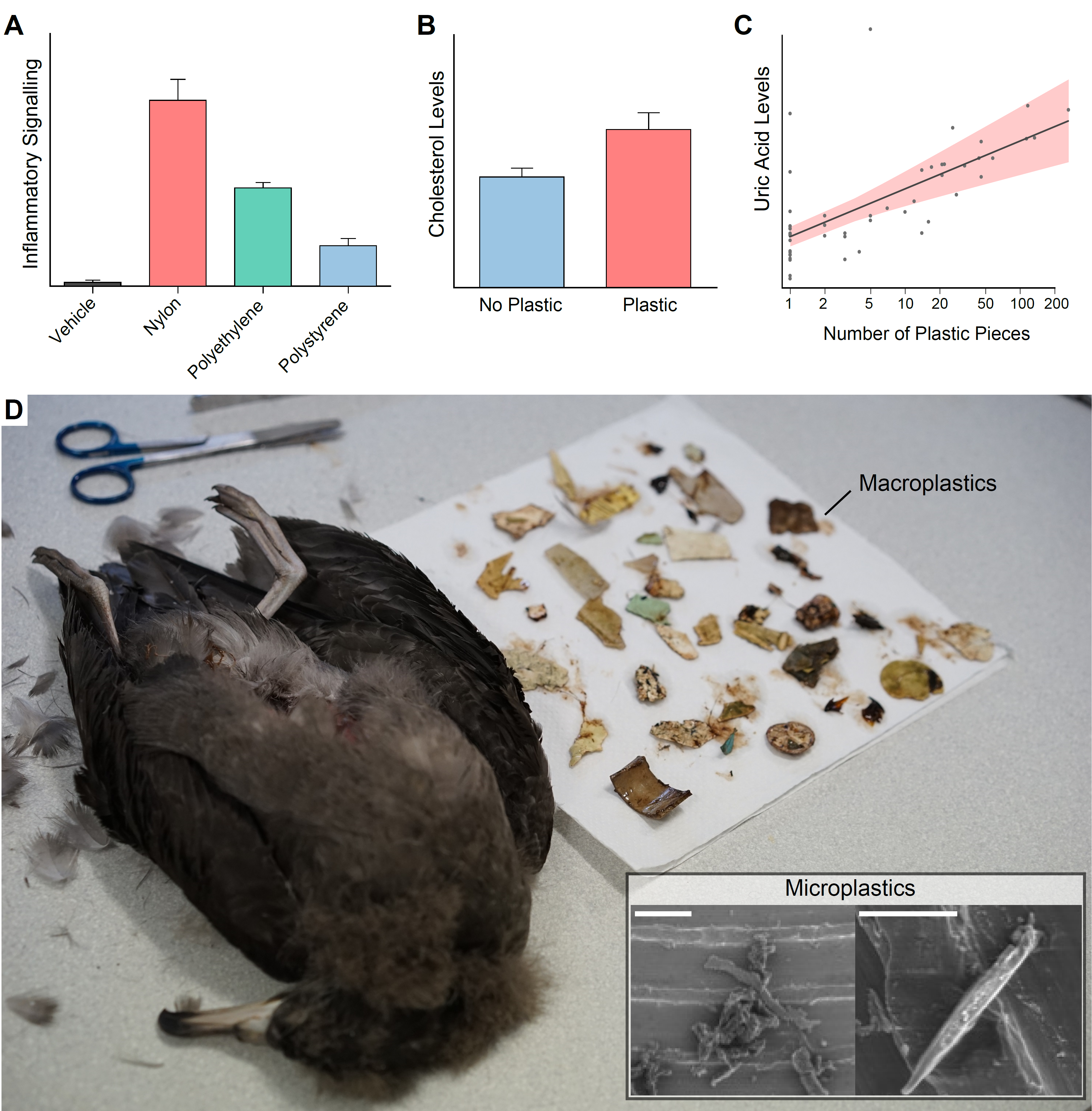
Figure. A) In cell culture models microplastics were inflammatory and this response differed between polymer composition. B and C) In seabirds, plastic ingestion was associated with significantly increased cholesterol (B) and uric acid (C) in the plasma. D) Seabirds fatally ingest macroplastics, but the sublethal effects of microplastics are unknown (Scale bars 10µm) (Lavers et al. 2014).

Epidemiological Research into Alzheimer’s Disease and Common Pain Relieving Drugs

Our understanding of the pathophysiological mechanisms of Alzheimer’s disease (AD) remains relatively unclear; however, the role of neuroinflammation as a key etiological feature is now widely accepted due to the consensus of epidemiological, neuroimaging, preclinical and genetic evidence. Consequently, non-steroidal anti-inflammatories (NSAIDs) have been investigated in epidemiological and clinical studies as potential disease modifying agents. Previous epidemiological studies focused on incidence of AD and did not thoroughly parse the effects at the individual drug level. The therapeutic potential of modifying incidence has a number of limitations, and we now know that each NSAID subtype has a unique profile of physiological impacts corresponding to different therapeutic profiles for AD. Therefore, we utilized the AD Neuroimaging Initiative (ADNI) dataset to investigate how the use of common NSAIDs and paracetamol alter cognitive decline in subjects with mild cognitive impairment (MCI) or AD.


Negative binomial generalized linear mixed modelling was utilized to model the cognitive decline of 1619 individuals from the ADNI dataset. Both the mini-mental state examination (MMSE) and Alzheimer’s disease assessment scale (ADAS) were investigated. Explanatory variables were included or excluded from the model in a stepwise fashion with Chi-square log-likelihood and Akaike information criteria used as selection criteria. Explanatory variables investigated were APOE4, age, diagnosis (control, MCI or AD), gender, education level, vascular pathology, diabetes and drug use (naproxen, celecoxib, diclofenac, aspirin, ibuprofen or paracetamol).


The NSAIDs, aspirin, ibuprofen, naproxen and celecoxib did not significantly alter cognitive decline. However, diclofenac use was associated with slower cognitive decline (ADAS χ2=4.0, p=0.0455, MMSE χ2=4.8, p=0.029). Paracetamol use was associated with accelerated decline (ADAS χ2=6.6, p=0.010, MMSE χ2=8.4, p=0.004). The APOE4 allele was associated with accelerated cognitive deterioration (ADAS χ2=316.0, p<0.0001, MMSE χ2=191.0, p<0.0001).


This study thoroughly investigated the effects of common NSAIDs and paracetamol use on cognitive decline in MCI and AD subjects. Most common NSAIDs did not alter cognitive decline. However, diclofenac use was associated with slowed cognitive deterioration, providing exciting evidence for a potential disease modifying therapeutic. Conversely, paracetamol use was associated with accelerated decline; which, if confirmed to be causative, would have massive ramifications for the recommended use of this prolific drug.

Zinc deficiency and Alzheimer’s disease

The risk of developing Alzheimer’s disease (AD) increases dramatically as we age. Similarly, the common malnutrition of zinc deficiency is also more prevalent in the elderly more due to changes in diet and zinc absorption as we age. We have shown that zinc deficiency induces the secretion of the inflammatory cytokine interleukin-1β (IL-1β) in macrophages and microglia. This is due to the activation of the IL-1β regulatory protein complex the NLRP3 inflammasome. Neuroinflammation has long been suspected as an exacerbating factor in AD pathology and recently the NLRP3 pattern recognition receptor (PRR) has been found to be the critical in regulated neuroinflammation in mouse models of AD. It is thought that AD associated toxic amyloid oligomers cause disruption in microglia physiology which initiates the formation of the multiprotein NLRP3 inflammasome complex causing the secretion of the IL-1 β. Therefore, this study aimed to assess whether a zinc deficient diet would accelerate the AD behavioural phenotype seen in the APPswe/PS1 mouse model of AD by inducing inflammation through NLRP3 activation. To test this hypothesis, mice were placed on a zinc deficient (3mg/kg) or a zinc normal (35mg/kg) diet for six months. Cognitive/memory performance was evaluated with the Morris water maze, Y-maze and novel smell tasks at baseline, three and six months on diet. To assess if the effect of zinc deficiency was reversible, at the 3 month time-point half the zinc deficient mice were placed back on a zinc normal diet. Our results suggest that zinc deficiency caused memory deficits in the APPswe/PS1 mice at 3 and 6 months on diet while at the same age the wild type mice on a zinc deficient diet and the APPswe/PS1 mice on normal diet showed no deficits. Furthermore, placing the zinc deficient mice on a zinc normal diet partially reverse the memory deficits. Additionally, it was found that innate immune cells from the zinc deficient mice had a hyper-inflammatory phenotype. Therefore, we have shown that zinc deficiency accelerates the AD phenotype in the APPswe/PS1 mice potentially through an inflammatory mechanism. This research aims to further investigate the role of zinc deficiency and NLRP3 in AD using NLRP3-/- mice and novel inhibitors of NLRP3 activation.

Cannabinoids

Cannabis sativa is an herbaceous plant that when consumed produces a number of well-known effects on the body, primarily elicited by two G-protein coupled receptors: cannabinoid receptor type 1 (CBI) and type 2 (CBII). Activation of the CBII receptor suppresses the inflammatory response of the immune system. Research has suggested that inflammation can be pathological in central nervous system (CNS) injuries and during early development the brain may be more sensitive to inflammation. Therefore, this research aimed to assess if CBII selective agonists could be neuroprotective in an animal model of childhood cerebral hypoxia (CCH) through an anti-inflammatory mechanism. A previously established model of CCH, that involved the ligation of the left carotid artery of a P26 rat, followed by a fixed hypoxic period, was chosen because of age suitability and its applicability to both asphyxia and stroke pathologies. Investigations into the variability of infarction in the established model, lead to the development of a new model that ended following a clonic tonic seizure. This new model caused significantly less variable brain infarctions compared to the previously reported model and was named Variable hypoxia ischemia (Variable HI). Two CBII agonists were then tested for neuroprotective properties in Variable HI using a partial agonist (GW405833) and a full agonist (HU910). Both single and multiple drug administration strategies were tested and found to provide no substantial tissue protection. Furthermore, behavioral tests were performed following the multiple administration strategy and no differences were found in any functional outcome. Therefore, the anti-inflammatory effects of CBII agonists are unlikely to be substantially therapeutic following CCH. It was argued that this lack of efficacy was caused by the possibility that inflammation is not truly pathological following CCH.

Morris Water Maze illustrated by Jack Rivers

Developing a New Animal Model

      During my research on early child asphyxia I found the rat model currently used in the literature produced highly variable injuries. After researching I hypothesized that the probable cause of these inconsistencies is the nature of the blood vessels in the brain. Like any blood vessel map they differ greatly between individuals. As an example, compare the blood vessels of the inside of your forearm to your friends, you’ll find that they cross in different places and potentially you may have some  vessels that your friend doesn’t, or vice versa. This variability occurs in the brain as well, in fact, in anatomy books you’ll see a ring of blood vessels in  the middle of your head called the “circle of Willis” this is supposed to operate like a back-up system. The circle has four major blood vessels -feeding  it (two carotid and two vertebral), the idea is that if one of these supplies fails, the brain can continue functioning as the circle of Willis that supplies  the brain will still have a rich supply of blood from the other three blood vessels. However, the anatomy books don’t tell you that this is only intact in some of the population; most of the population have a semi-circle.

      This variability of the circle of Willis is similar in the rat. In the model for early childhood asphyxia one of these supplies to the circle of Willis is tied off and then the rat is put in a low oxygen environment for 60 minutes. However, if some rats have a complete circle of Willis and some rats do not, the injury could be different and as my research found out, this difference  could be huge! So I developed a model where instead of placing the rat in the low oxygen environment for 60 minutes, we placed each rat in low oxygen and  observed the behaviour of the rat, we only removed the rat from the low oxygen once we saw a particular behaviour. This new model produced a more consistent  brain injury. This meant we could use fewer rats to do the same experiment, reducing experimental cost and reducing the amount of rats needed, which helps  me sleep at night.

To find out more, read my journal article published in Journal of Neuroscience Methods.

Download (PDF)

Blood Vessel ligation. Illustrated by Jack Rivers

Hydrogen Sulfide in Inflammation

     Sepsis is the pathological presence of an infection in the blood or other tissue; it is commonly caused by bacterial infection and can lead to multiple organ failure and ultimately death. This area is of ever increasing importance as more and more cases of antibiotic resistance infections occur. However, in many cases it is not the infection that directly kills the patient; it is an uncontrolled overreaction by the immune system. This pathological immune system response is referred to as systemic inflammatory response syndrome (SIRS) and is characterized by hypotension, tachycardia and a higher or lower body temperature. SIRS can result in multi-organ failure, particularly damage to the lungs with acute respiratory distress syndrome. Our research aims to develop effective techniques for quickly diagnosing sepsis and SIRS using novel biomarkers, as well as developing new treatments using a combination of cellular models of the disease and clinical samples from sepsis sufferers.

     The immune system’s response to infection and toxins is a normal and crucial function of human physiology. Upon recognition of an infection or toxin, immune cells produce a range of signaling molecules including cytokines and chemokines to further recruit and activate immune cells. However, the immune system can produce cytokines and chemokines at cytotoxic levels, as well as other cytotoxic compounds such as reactive oxygen species (ROS) including free radicals and reactive nitrogen species (RNS) such as nitric oxide.  In sepsis induced systemic inflammatory response syndrome (SIRS), the cytotoxic compounds can reach dangerous levels in the blood, causing damage to sensitive organs such as the lungs and the kidneys. The downstream effects of this organ damage, such as hypotension and respiratory failure, can result in further organ failure and death. Two molecules that may be crucial in this pathological activation of immune cells are hydrogen sulfide and substance P.

      Hydrogen sulfide is a novel gaseous signaling molecule, at normal physiological levels it is crucial in many processes ranging from memory and learning to vasodilatation.  It is produced at high levels during inflammation, this signals the recruitment and activation of immune cells. Hydrogen sulfide is produced through the removal of the thiol group of L-cysteine by the enzymes cystathionine γ lyase (CSE) systemically or cystathionine β synthase (CBS) in the central nervous system. The exact mechanisms of how hydrogen sulfide elicits its effects are still being elucidated, however, it is clear that it involves the modulation of cellular function through MAP/ERK signaling pathways, vanilloid receptor pathways, and possibly through altering cellular glutathione oxidation states. Animal models of sepsis have shown that reducing the levels of hydrogen sulfide production by administering CSE enzyme inhibitors, improved the animals’ condition and increased survival rates.

      Substance P is a small peptide that is a signaling molecule involved in inflammation and pain. It is produced by afferent somatic sensory neurons and activated macrophages and causes an increase in immune cell activation, vasodialation and pain signaling. Substance P acts, at least in part, through binding to the neurokinin 1 (NK-1) receptor causing cellular changes through the MAP/ERK pathways to illicit its effects.  Nk-1 antagonists have been shown in animal models of sepsis to be protective of lung injury, demonstrating the importance of substance P in sepsis induced SIRS.

      Because hydrogen sulfide and substance P are involved in pathological immune cell activation, there is potential for them to be used as biomarkers of sepsis and sepsis induced SIRS. They are also an attractive potential target of pharmacological intervention for the treatment of inflammatory diseases such as SIRS.

Antibody Specificity

      Immunohistochemistry is a specific protein stain that is used regularly throughout scientific research. Immunohistochemistry is a tissue stain that uses the specific binding of antibodies produced when an epitope (antigen or small segment of protein) is exposed to the immune cells of an animal from a different species. It involves the purification of a segment of a specific protein (epitope) that is to be investigated. This epitope is then injected to an animal that is a different species to the epitope host animal. After injection the animal will have an immune response to the epitope and will produce antibodies that will bind to the epitope specifically. These antibodies (referred to as the primary antibody) are then extracted from the animal’s serum and applied to the tissue that is to be stained for the epitope. The primary antibody solution is then washed off, leaving only the primary antibodies that are bound to the epitope on the tissue. The primary antibody will then be visualized by a number of methods.

      Problems occur when these antibodies bind to many proteins non-specifically. This can happen in a number of ways (see figure below), this leads to a miss-leading stain. There is huge debate in the literature about where the CBII receptor is, this is due largely to the unreliability of immunohistochemistry. We performed a study with 4 separate commercially bought CBII antibodies and one privately produced antibody. Using these we stained neurologically injured brains and found that one antibody showed large about of CBII expression on the injured side of the brain, another antibody showed only a small localized patch of staining right near the injury, yet another showed CBII expression over the entire brain, and the other two showed now staining at all. This demonstrated clearly the difficulties of immunohistochemistry and illustrated the need for the use of more than one technique (like in situ hybridization) and more than one antibody.

Ways in which labeling can occur in the tissue following immunohistochemistry. A- specific immunohistochemistry labeling of a target GRPC protein using primary antibody with a two biotinylated secondary antibodies attach, avidin has bound to the biotin on the secondary and three HRP conjugated biotins have bound to the avidins, the HRP has then facilitated the oxidation of colourless and soluble DAB into a darkened insoluble form. B- Nonspecific binding of the primary antibody to proteins on or within the cell, this can be binding of the binding domain (left) or binding of another region of the antibody (right). C- Nonspecific binding of the secondary antibody to proteins on or within the cell, this can be binding of the binding domain (left) or bind of another region of the antibody (right). D. Nonspecific binding of biotin (left) and avidin (right) to proteins within the tissue. E. Endogenous peroxidase activity that can oxidize DAB, this can be facilitated by membrane bound or free floating peroxidases. Illustrated by Jack Rivers